Gene mutations
The word mutation means a change in the genetic composition of a cell. Mutations can be divided into two main groups. The first group is gene mutations. These are chemical changes in the DNA of the cell.
A gene mutation is a change in the order of bases on a strand of DNA.
Section of a DNA molecule
The word mutation means a change in the genetic composition of a cell. Mutations can be divided into two main groups. The first group is gene mutations. These are chemical changes in the DNA of the cell.
A gene mutation is a change in the order of bases on a strand of DNA.
Section of a DNA molecule
Gene mutations
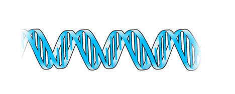
DNA exists as two helical strands held together through the base pairs by hydrogen bonding.
Gene Mutations
Gene Mutations
Original gene structure
Original gene structure
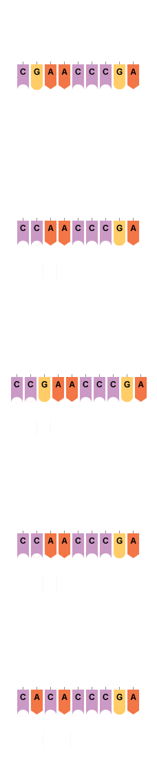
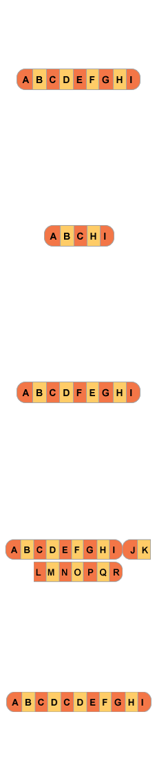
This shows the normal order of the bases for one of the strands in the area highlighted in the DNA molecule. Only one of the strands of DNA is involved in protein synthesis. A change from the normal order of bases leads to different types of gene mutation.
Here you can see the strand after one nucleotide pair is substituted by another nucleotide pair in the strand of DNA. This is called substitution. The first of the nucleotides with base G has been substituted for a nucleotide with base C. Any one of the nucleotide pairs can be substituted.
Here you can see the strand after one pair of nucleotides is inserted into the strand of DNA. This is called insertion. A nucleotide with base G is inserted. More than one pair of nucleotides can be inserted.
Here you see the strand after one pair of nucleotides is removed from the strand of DNA. This is called deletion. The nucleotide with the base G has been removed. More than one pair of nucleotides can be deleted.
Here you can see the strand after two pairs of nucleotides break off from the strand of DNA and rotate before rejoining into the strand. This is called inversion. The first pair of nucleotides on the strand with bases C and A has inverted and is now shown as bases A and C. More than two pairs of nucleotides can be inverted.
Sickle Cell Anaemia
Sickle Cell Anaemia
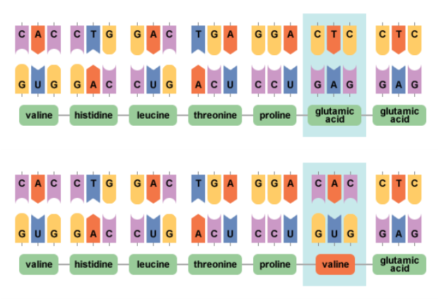
The diagram above shows the difference in the synthesis of haemoglobin in normal DNA and abnormal DNA. The top strand shows the normal DNA, mRNA and amino acid sequences. A single substitution mutation has changed the base sequence in the DNA. The base sequence on the mRNA produced by the DNA is altered. As a result, a codon on the mRNA is altered. A different amino acid is inserted into the protein chain. The amino acid valine is inserted instead of glutamic acid, which is shown by the bottom strand of DNA. The protein synthesised is altered and no longer functions normally. These sequences result in sickle-shaped red blood cells. This blood disorder is known as sickle cell anaemia.
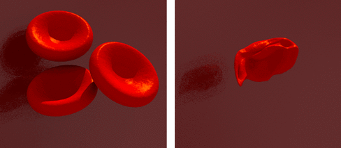
red blood cells
Phenylketonuria
Phenylketonuria
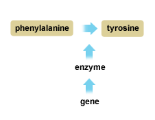
The diagram represents part of the normal metabolic pathway involving the amino acid phenylalanine within human cells. The gene controls the synthesis of the enzyme and the enzyme converts the amino acid phenylalanine to tyrosine.
If a gene mutation occurs then individuals with this mutation can no longer produce the normal enzyme. This in turn means that they can no longer convert phenylalanine to tyrosine. The failure to bring about the normal enzyme conversion is called a metabolic block.
If a gene mutation occurs then individuals with this mutation can no longer produce the normal enzyme. This in turn means that they can no longer convert phenylalanine to tyrosine. The failure to bring about the normal enzyme conversion is called a metabolic block.
In newly born babies this metabolic block leads to the condition phenylketonuria (PKU). The effects of the condition if undetected and treated at birth are that:
- phenylalanine concentration builds up within the body of the new born baby
- phenylaline is converted into toxic substances
- the toxic substances cause irreversible damage to the developing brain
Changes in the structure of the chromosome
Chromosome mutations
Chromosome mutations are due to change in either the chromosome structure or the chromosome number of a cell.
During Meiosis chiasmata form and pieces of chromatid from homologous pairs of chromosomes may break off and crossover. This gives new combinations of the different alleles of the different genes. Failure in normal crossing-over can lead to the following chromosome mutations:
During Meiosis chiasmata form and pieces of chromatid from homologous pairs of chromosomes may break off and crossover. This gives new combinations of the different alleles of the different genes. Failure in normal crossing-over can lead to the following chromosome mutations:
Normal chromosome structure
Normal chromosome structure
Here you can see the normal chromosome structure.
Here you can see deletion. This is where the genes of a chromosome are lost. Inherited chromosome mutations such as deletion may be lethal.
Here you can see inversion. This is where the order of the genes on the chromosome is changed.
Here you can see the process of translocation. This is when genes from one chromosome break off and bind to a different chromosome. Inherited chromosome mutations such as translocation may be lethal.
Here you can see duplication. This is when genes are copied and inserted into the chromosome.
Non-disjunction and mutagenic agents
Non-disjunction
Non-disjunction
Non-disjunction
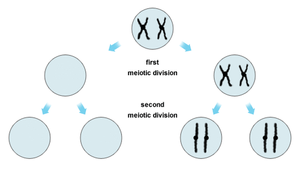
During meiosis, when two homologous chromosomes, instead of separating, both go into the same gamete. This is known as non-disjunction. As a result of non-disjunction some gametes have two copies of a chromosome and others lack a copy. The diagram shows only one pair of homologous chromosomes as they would align at the start of meiosis. (In the cell of a human female 23 homologous pairs would align). After the first meiotic division the homologous pair, instead of separating, have both remained in the same cell. After the second meiotic division the gametes formed are of two types; half lack a chromosome and half have two copies of the same chromosome.
Down's Syndrome
In humans, if an ovum with two copies of chromosome 21 is fertilised by a normal sperm cell then the individual formed has three copies of chromosome 21 in their cells. Such individuals show Down's syndrome.
Down's Syndrome
In humans, if an ovum with two copies of chromosome 21 is fertilised by a normal sperm cell then the individual formed has three copies of chromosome 21 in their cells. Such individuals show Down's syndrome.
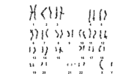
karyotype
A karyotype shows the full set of chromosomes of an individual arranged in sequence. Here the karyotype shown is from the cell of a male with Down's syndrome. Chromosomes one to 20 and 22 are shown to be present as homologous pairs. There are three copies of chromosome 21 and such individuals develop to show Down's syndrome. The sex chromosomes are shown as X and Y and such individuals develop as male.
Mutagenic agents
Gene and chromosome mutations occur naturally and generally have a low frequency. However, certain chemicals and rays can increase the rate of frequency of mutation. These are called mutagenic agents and include:
Gene and chromosome mutations occur naturally and generally have a low frequency. However, certain chemicals and rays can increase the rate of frequency of mutation. These are called mutagenic agents and include:
- chemicals such as mustard gas, caffeine and colchicine
- irradiation such as U-V radiation, X-rays and gamma rays
PolyploidyPolyploidy is a condition in which an individual has sets of chromosomes greater than the normal diploid number.
There are number of key factors to help you remember:
There are number of key factors to help you remember:
- The number of chromosomes making up one set is the haploid number = n.
- The number of chromosomes making up two sets is the diploid number = 2n.
- Polyploid species would be represented by 3n, 4n, etc.
- Polyploids with even numbers of sets of chromosomes are fertile as the chromosomes can form as homologous pairs in meiosis.
- Polyploids with uneven numbers of sets of chromosomes are sterile as the chromosomes are unable to form as homologous pairs in meiosis.
Polyploid formation
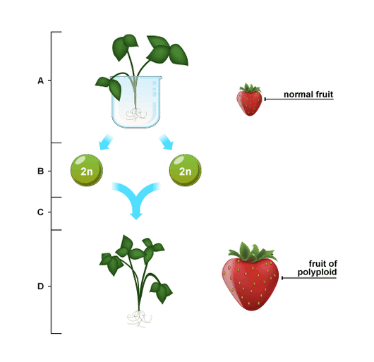
Polyploid formation
At A the strawberry plant is exposed to a chemical called colchicines. At B due to the effect of colchicines, total non-disjunction at meiosis produces diploid gametes (2n). As a result, at C two diploid gametes fuse at fertilisation. Finally at D a new plant develops which has four sets of chromosomes (4n).
Closely related plant species can be crossed to create polyploids. These plants show hybrid vigour. Man makes use of increased hybrid vigour to develop better crop plants. Hybrid vigour is shown by polyploids through higher yield of fruit and greater resistance to disease and drought.
Closely related plant species can be crossed to create polyploids. These plants show hybrid vigour. Man makes use of increased hybrid vigour to develop better crop plants. Hybrid vigour is shown by polyploids through higher yield of fruit and greater resistance to disease and drought.
